Your Guide to Dental X-Ray Safety: A Checklist Template for Radiation Protection & Calibration
Published: 09/10/2025 Updated: 12/13/2025
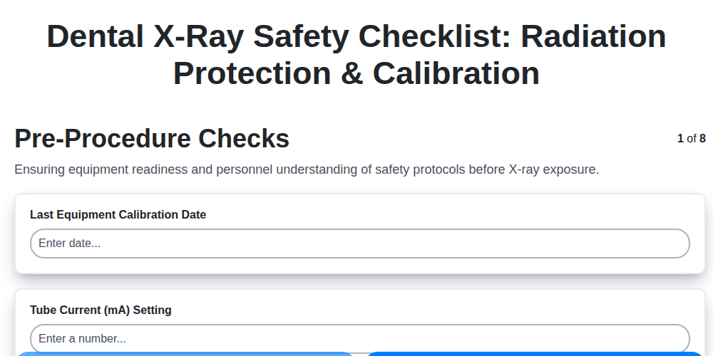
Table of Contents
- Why Dental X-Ray Safety Matters
- Understanding Your Responsibilities
- Pre-Procedure Checklist: Patient Screening & Preparation
- Equipment Calibration & Testing: Ensuring Accuracy
- Shielding & Protective Barriers: Minimizing Exposure
- Exposure Parameters: Optimizing Image Quality & Dose
- Personnel Safety & Monitoring: Protecting Your Team
- Record Keeping & Documentation: Maintaining Compliance
- Emergency Procedures: A Plan for Unexpected Events
- Resources & Links
TLDR: Worried about dental x-ray safety? Our free checklist template makes it easy to ensure patient and staff protection. It covers everything from equipment calibration and shielding to personnel monitoring and emergency procedures - a simple way to stay compliant and minimize radiation risk. Download it now for a safer dental practice!
Why Dental X-Ray Safety Matters
The health and well-being of your patients and team are paramount. While dental X-rays offer invaluable diagnostic information, they involve exposure to ionizing radiation-a potentially harmful element if not managed responsibly. Minimizing this risk isn't just about adhering to regulations; it's about upholding a commitment to ethical practice and patient-centered care.
Overexposure to radiation, even in small amounts, can contribute to an increased risk of long-term health problems. For patients, this can manifest as a heightened susceptibility to certain cancers. For dental professionals, consistent exposure without proper protection can lead to cumulative health risks.
Beyond the direct health implications, a strong safety protocol fosters trust and confidence. Patients appreciate knowing that their dental practice prioritizes their well-being and takes proactive measures to safeguard their health. Similarly, a culture of safety enhances team morale and reinforces a shared commitment to excellence. Ultimately, prioritizing dental X-ray safety isn't just about following procedures - it's about building a practice based on trust, responsibility, and the highest standards of care.
Understanding Your Responsibilities
As a dental professional, your commitment to patient and staff safety goes beyond simply possessing the necessary equipment. It's a legal and ethical obligation rooted in the potential risks associated with ionizing radiation. Understanding these responsibilities is paramount to maintaining a safe and compliant practice.
Your primary duty is to minimize radiation exposure to both patients and personnel-the ALARA (As Low As Reasonably Achievable) principle dictates this. This involves more than just following procedures; it necessitates a proactive and informed approach. You're responsible for ensuring all team members are adequately trained and understand their roles in radiation safety protocols. Maintaining accurate records of equipment calibration, patient exposure, and personnel dosimetry is crucial-these serve as a tangible demonstration of your commitment and provide a vital audit trail. Furthermore, you must remain updated on evolving regulations and best practices, actively seeking professional development opportunities to refine your safety protocols. Finally, open communication with patients about the risks and benefits of radiographic procedures is essential to informed consent and fostering a trusting relationship. A conscientious approach to these responsibilities not only safeguards those in your care but also reinforces the integrity of your practice.
Pre-Procedure Checklist: Patient Screening & Preparation
Before initiating any dental radiographic procedure, a thorough patient screening and preparation phase is crucial for minimizing radiation exposure and maximizing image quality. This involves more than just a quick question; it demands a systematic approach to ensure patient safety and optimize the radiographic process.
Radiographic History & Justification: Begin by meticulously reviewing the patient's radiographic history. Determine the dates of previous films, the areas examined, and the reasons for the initial radiographs. This assessment helps avoid unnecessary repetition and justifies the need for new exposures. Clearly documenting this rationale is vital for demonstrating adherence to ALARA principles.
Pregnancy Screening: A Non-Negotiable Step: A robust and documented pregnancy screening protocol is paramount. Directly and sensitively inquire about the possibility of pregnancy in all female patients of childbearing age. Clearly explain the potential risks of radiation exposure during pregnancy and offer alternative diagnostic methods if concerns arise. Record the inquiry and the patient's response in the patient's file. Remember, even low doses of radiation can pose a risk to a developing fetus.
Explain the Procedure & Gain Informed Consent: Communicate clearly and concisely the purpose of the radiographic examination, the type of images to be taken, and the anticipated radiation exposure. Address any patient questions or concerns openly and honestly. While not always a formal "consent form," the patient's understanding and agreement are essential.
Patient Positioning Considerations: Optimal patient positioning not only improves image quality but also reduces the need for retakes, thereby minimizing radiation exposure. Ensure the patient is properly positioned and stabilized to avoid movement during the exposure.
Proper patient preparation minimizes exposure and ensures diagnostic images.
Equipment Calibration & Testing: Ensuring Accuracy
Maintaining the accuracy of your dental x-ray equipment is paramount to both patient safety and diagnostic quality. Regular calibration and testing aren't merely best practices-they're regulatory requirements. A malfunctioning machine can lead to unnecessary patient exposure, poor image quality requiring retakes, and potential legal liabilities.
Here's a breakdown of essential calibration and testing procedures:
1. Timer Accuracy Verification: The timer dictates exposure duration. Deviations, even minor ones, can significantly impact radiation dose. Use a calibrated timer or a known accurate time source to check timer accuracy at least annually, and whenever equipment is serviced. A variance of more than ±5% warrants immediate correction.
2. Tube Current (mA) and Voltage (kVp) Verification: Utilize a dedicated reference dosimeter or a calibrated ionization chamber to verify the actual output of the x-ray tube. Confirm that the machine's settings accurately reflect the delivered values.
3. Collimation Check: Proper collimation restricts the radiation beam to the area of interest, minimizing scatter radiation and patient dose. Use a radiation survey meter to confirm that the collimation is accurate and effective. A visible light collimation check can provide a visual confirmation.
4. Leakage Radiation Testing: This is a critical annual test. Use a calibrated survey meter to measure leakage radiation emanating from the x-ray tube housing. Readings must be below regulatory limits, typically very low (e.g., 0.1 mR/hr at 1 meter). Any detectable leakage requires immediate repair or replacement.
5. Image Receptor Testing (Digital Sensors & CR Plates): Regularly assess the functionality and sensitivity of your digital sensors and CR plates. This includes checking for uniformity, latitude, and detective quantum efficiency (DQE). Software integrated into digital radiography systems often provides automated testing capabilities.
Record Keeping is Key: Meticulously document all calibration and testing results. Keep these records readily available for review by regulatory agencies and your Radiation Safety Officer. Frequency and scope of testing should be determined by your RSO and aligned with manufacturer recommendations and regulatory requirements.
Shielding & Protective Barriers: Minimizing Exposure
Effective shielding and protective barriers are your first line of defense against scattered radiation. While collimation minimizes the primary beam, scattered radiation can still pose a risk to both patients and personnel. Here's what you need to know to create a safe radiographic environment:
Lead Aprons & Thyroid Shields: The Essentials
Lead aprons are non-negotiable for patients and anyone in the room during an exposure. Ensure they are appropriately sized, cover the entire abdomen and pelvis, and include a thyroid shield to protect the thyroid gland - a particularly vulnerable area. Regularly inspect these shields for cracks, holes, or signs of degradation, as damage significantly reduces their effectiveness. Replacement is crucial for compromised shields.
Room Shielding: Walls and Doors
Many modern dental offices incorporate lead shielding into the walls and doors of the x-ray room. This is particularly important for rooms where multiple exposures are performed. If your room lacks built-in shielding, consider the possibility of adding it, consulting with a qualified contractor.
Distance: Your Best Friend
Remember that radiation intensity decreases dramatically with distance. Increasing the distance between personnel and the x-ray beam provides significant protection. Implement procedures to ensure operators and assistants maintain a safe distance whenever possible.
Dose Area Product (DAP) Monitoring (Where Applicable)
Some practices utilize DAP meters to track the total radiation dose delivered during a period. This data can help identify areas for improvement in technique and workflow to minimize overall exposure.
Regular Inspections
Conduct routine visual inspections of all shielding and protective barriers to confirm their integrity and effectiveness. Document these inspections meticulously in your quality assurance records.
Exposure Parameters: Optimizing Image Quality & Dose
Achieving high-quality radiographic images while minimizing patient exposure requires a deep understanding and precise application of exposure parameters. It's a balancing act - too little radiation, and you get a noisy, unreadable image; too much, and you unnecessarily increase the patient's radiation dose. Here's a breakdown of the key factors and how to optimize them:
Kilovoltage (kVp): The Energy Level
kVp primarily affects image contrast. Higher kVp values penetrate tissues more effectively, reducing contrast but decreasing dose. Lower kVp values increase contrast but require more mAs to penetrate, potentially raising the dose. For most intraoral radiographs, a kVp range of 60-80 is typical. Consider the anatomy being imaged; thicker structures may require slightly higher kVp.
Milliamperage-seconds (mAs): Quantity of Radiation
mAs directly influences the number of X-ray photons reaching the image receptor, thus impacting image density. A higher mAs value results in a denser image. It's crucial to select the lowest mAs that still produces an acceptable image. Digital radiography allows for greater latitude in mAs selection; however, always prioritize ALARA principles.
Distance (SID): A Critical Factor
The source-to-image distance (SID) is a key determinant of dose. Increasing the SID reduces the dose received by the patient. The standard SID for most dental radiography is 40 inches (100 cm). Always maintain a consistent SID for repeatable results.
Filtration:
Filtration reduces the low-energy X-ray photons that contribute to skin dose but don't contribute to the image. Proper filtration is essential for minimizing patient exposure, especially for pediatric patients.
Technique Charts & Digital Latitude:
Utilize technique charts as a starting point, but always adapt based on patient size, tissue density, and radiographic requirements. Digital radiography offers considerable latitude, allowing for adjustments without significantly compromising image quality. However, resist the temptation to excessively increase mAs, as this directly increases radiation dose. Carefully evaluate the image at the lowest possible mAs setting.
Personnel Safety & Monitoring: Protecting Your Team
Radiation safety isn't just about protecting patients; it's paramount for safeguarding the well-being of your dental team. Consistent exposure, even at seemingly low levels, can accumulate over time and pose health risks. A robust personnel safety and monitoring program is therefore non-negotiable.
Here's what you need to prioritize:
- Personal Dosimetry: All personnel who potentially receive radiation exposure - including dentists, dental hygienists, dental assistants, and even maintenance staff - must wear and properly utilize personal dosimeters. These badges (either film badges or electronic personal dosimeters) accurately measure cumulative radiation exposure. Regularly review dosimetry reports and address any exceedances immediately.
- ALARA (As Low As Reasonably Achievable): Embrace the ALARA principle in every aspect of your practice. This means making conscious efforts to minimize exposure whenever possible. Consider techniques like:
- Optimizing radiographic positioning to reduce retakes.
- Utilizing proper shielding techniques.
- Limiting exposure time.
- Maintaining a safe distance from the primary beam.
- Comprehensive Training: Ensure all team members receive thorough and ongoing radiation safety training. This training should cover topics like:
- Radiation physics basics
- Safe radiographic techniques
- Proper use of personal protective equipment (PPE)
- Emergency procedures
- Shielding and Protective Barriers: Always utilize lead aprons and thyroid shields when performing radiographic procedures. Regularly inspect these protective devices for damage and replace them as needed.
- Awareness and Communication: Foster a culture of safety where team members feel comfortable raising concerns and reporting potential radiation exposures. Open communication is vital for continuous improvement.
Record Keeping & Documentation: Maintaining Compliance
Maintaining meticulous records is far more than just good practice; it's a cornerstone of compliance and a vital shield against potential liability. A robust record-keeping system demonstrates a proactive commitment to patient and personnel safety, providing evidence of due diligence in the event of an audit or incident.
What should you be documenting? It goes well beyond just exposure logs. Think comprehensively:
- Exposure Logs: Each radiograph requires a log entry detailing patient name, date, type of procedure, technique factors (kVp, mA, time, SID), and the name/title of the operator.
- Calibration Records: Maintain detailed records of all equipment calibrations and acceptance tests, including dates, results, and the name of the individual performing the tests. These records must be readily available for review.
- Personnel Dosimetry Records: Securely store individual personnel dosimetry reports (film badges or electronic dosimeters), tracking cumulative exposure over time. These records are legally protected and require specific handling procedures.
- Quality Assurance (QA) Reports: Document regular QA activities, outlining any findings, corrective actions taken, and verification of those actions.
- Equipment Maintenance & Repair Logs: Keep detailed records of all maintenance, repairs, and modifications made to the radiographic equipment.
- Training Records: Document all radiation safety training received by personnel, including dates, topics covered, and assessments.
- Incident Reports: In the unfortunate event of a radiation incident, a thorough report detailing the event, actions taken, and corrective measures implemented is crucial.
Retention Periods: Be aware that legal requirements dictate specific retention periods for these records. Consult with your RSO and legal counsel to ensure you are complying with all applicable regulations. Electronic record-keeping systems are increasingly common, offering significant benefits for organization, accessibility, and security - but ensure they meet regulatory requirements for data integrity and security.
Emergency Procedures: A Plan for Unexpected Events
Even with the most rigorous safety protocols in place, unexpected events can occur. A well-defined emergency response plan is crucial for minimizing risk and ensuring the safety of patients and personnel. This isn't just about theoretical preparation; it's about having a clear, actionable plan to follow when things go wrong.
Common Emergency Scenarios & Response
Let's outline some potential scenarios and the steps to take:
- Equipment Malfunction During Exposure: Immediately halt the exposure. Instruct the patient and any nearby personnel to leave the room. Isolate the equipment and contact a qualified service technician for repair and assessment. Document the incident thoroughly.
- Leakage Radiation Detection: If a leakage radiation survey detects an unsafe level of radiation, immediately shut down the x-ray equipment and contact the Radiation Safety Officer (RSO). Restrict access to the area until the problem is resolved and cleared by a qualified technician.
- Personnel Overexposure (Suspected): If you suspect a personnel member has received an excessive dose of radiation, immediately remove them from the area. Follow established protocols for reporting the incident to the RSO and relevant authorities. Medical consultation is mandatory.
- Fire in the X-Ray Room: Follow established fire safety procedures, including activating the fire alarm, evacuating the area, and contacting emergency services. X-ray equipment must be de-energized if safe to do so.
- Unresponsive Patient: If a patient becomes unresponsive during or after an exposure, immediately assess their condition and administer appropriate first aid. Call emergency medical services (EMS).
Key Components of an Effective Emergency Plan
- Clear Communication: Establish a system for clear and concise communication during an emergency.
- Designated Roles: Assign specific roles and responsibilities to personnel. Who is responsible for evacuating the area? Who calls emergency services?
- Emergency Contact List: Post a readily accessible list of emergency contacts, including the RSO, local emergency services, and equipment service technicians.
- Regular Drills: Conduct regular emergency drills to familiarize personnel with procedures and identify areas for improvement.
- Plan Review & Updates: Review and update the emergency plan at least annually, or more frequently if there are changes in personnel, equipment, or regulations.
Remember: Prevention is the best approach. Consistent adherence to safety protocols and regular equipment maintenance significantly reduce the likelihood of emergencies occurring.
Resources & Links
- U.S. Food and Drug Administration (FDA) - Dental Radiation : The FDA's website provides comprehensive information on dental radiation, including regulations, safety guidelines, and updates on technology. Crucial for understanding the legal and regulatory framework surrounding dental x-rays. Focuses on device approval and safety standards.
- American Dental Association (ADA) : The ADA offers resources for dental professionals, including guidelines, best practices, and continuing education courses related to radiation safety. Their publications and resources are essential for staying current on recommended procedures.
- American Society of Radiologic Technologists (ASRT) : ASRT provides education, resources, and advocacy for radiologic technologists. Their content addresses radiation safety practices and ethical considerations, vital for personnel training and understanding exposure limits.
- Occupational Safety and Health Administration (OSHA) : OSHA's website contains information regarding worker safety regulations, including those related to radiation exposure. Important for understanding legal responsibilities for protecting dental staff.
- Conference of Radiation Control Program Directors (CRCPD) : CRCPD is an organization for state radiation control program directors. Their website often provides links to state-specific radiation control regulations, essential for ensuring compliance with local laws (highly relevant for 'Record Keeping & Documentation' section).
- International Academy of Oral Medicine and Toxicology (IAOMT) : While their focus is broader, IAOMT resources often discuss concerns regarding low-level radiation exposure and may be helpful for understanding the context of minimizing risk (useful for 'Why Dental X-Ray Safety Matters'). Approach with critical evaluation.
- National Institute of Standards and Technology (NIST) : NIST develops standards and technologies related to radiation measurement and calibration. Their resources may be relevant for the 'Equipment Calibration & Testing' section, although primarily for advanced understanding.
- Radcal - Dental X-ray Testing Equipment : Radcal specializes in equipment for testing and calibrating dental x-ray machines. Their website has informative content regarding calibration procedures and standards. Useful for deeper dive into 'Equipment Calibration & Testing'.
- H.P. Associates, Inc. : HPA provides radiation survey meters and consultation services. Their website offers information on radiation monitoring and measurement, valuable for 'Personnel Safety & Monitoring'.
- Therapy Services, Inc. : Therapy Services provides training, documentation templates, and support for dental practices regarding radiation safety. They offer practical tools for maintaining compliance and improving safety protocols - especially useful for the checklist.
Frequently Asked Questions
Who is responsible for ensuring dental X-ray safety?
Responsibility is shared. The dentist is ultimately responsible for overseeing patient safety and ensuring protocols are followed. The practice manager ensures procedures are in place and equipment is maintained. All staff members are responsible for adhering to safety protocols and reporting any concerns.
Found this Article helpful?
Dental Management Solution Demo
Simplify your dental practice & enhance patient care! ChecklistGuro streamlines appointment scheduling, patient records, and billing. Improve efficiency, reduce errors, & boost patient satisfaction. Manage it all with our Work OS.
Related Articles
Your Dental Office Safety Net: The Compliance & Patient Safety Checklist Template
Mastering Dental Recalls: Your Ultimate Checklist Template

Mastering Dental Case Presentations: Your Checklist Template for Success

Your Dental Compliance Checklist: A State & Federal Guide
Dental Operations Checklist: Boost Efficiency & Manage Risk

The Ultimate Dental Lab Case Checklist: Fabrication & Quality Assurance

Your Complete Dental Infection Control Checklist: PPE & Surface Disinfection
Mastering Dental Exams: Your Complete Charting Checklist Template
We can do it Together
Need help with
Dental Management?
Have a question? We're here to help. Please submit your inquiry, and we'll respond promptly.